 Skip to content
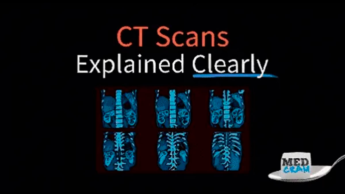
Nov
In the realm of medical imaging, computed tomography (CT) scans and high-resolution CT (HRCT) scans serve as invaluable tools for diagnosing and monitoring various health conditions. While both techniques utilize X-rays to create detailed images of the body’s internal structures, they differ significantly in their applications, imaging techniques, and the level of detail they provide. This article aims to clarify these differences and help you understand when each type of scan is used.
What is a CT Scan?
A CT scan, or computed tomography scan, is a diagnostic imaging procedure that combines X-ray images taken from different angles and uses computer processing to create cross-sectional images of bones, blood vessels, and soft tissues inside the body. CT scans are widely used for a variety of medical conditions, including:
- Trauma Assessment: Quickly identifying injuries in emergency situations.
- Tumor Detection: Locating and assessing the size of tumors in various organs.
- Internal Bleeding: Identifying sources of bleeding within the body.
- Infection Diagnosis: Detecting abscesses or other infections.
CT scans are typically performed with a standard resolution, which is sufficient for most diagnostic purposes. The images produced provide a comprehensive view of the anatomy, allowing physicians to make informed decisions regarding treatment.
What is a High-Resolution CT Scan (HRCT)?
High-Resolution CT (HRCT) is a specialized form of CT imaging that focuses on producing highly detailed images of the lungs and other structures. Unlike standard CT scans, HRCT employs thinner slices and a higher level of detail, making it particularly useful for evaluating lung diseases. HRCT is often used to diagnose conditions such as:
- Interstitial Lung Disease: Including pulmonary fibrosis and sarcoidosis.
- Chronic Obstructive Pulmonary Disease (COPD): Assessing the extent of airway obstruction.
- Lung Nodules: Characterizing and monitoring lung nodules for potential malignancy.
- Pneumonia: Providing detailed images to assess the severity and extent of lung infections.
The primary advantage of HRCT is its ability to reveal subtle changes in lung architecture that may not be visible on standard CT scans. This high level of detail is crucial for early diagnosis and management of various pulmonary conditions.
Key Differences Between CT and HRCT
- Image Resolution: The most significant difference lies in the resolution of the images produced. HRCT scans utilize thinner slices (typically 1-2 mm) compared to standard CT scans (usually 5-10 mm), allowing for a more detailed view of the structures being examined.
- Radiation Dose: HRCT scans often involve a higher radiation dose due to the need for multiple thin slices. However, advancements in technology have led to techniques that minimize radiation exposure while still providing high-quality images.
- Clinical Applications: While CT scans are versatile and used for a wide range of diagnostic purposes, HRCT is specifically tailored for lung evaluation. It is particularly beneficial in identifying and characterizing lung diseases that require a detailed assessment of the lung parenchyma.
- Imaging Technique: HRCT scans may employ specific techniques such as high-frequency algorithms and specialized reconstruction methods to enhance image quality, focusing on the lung fields and minimizing artifacts.
Conclusion
Both CT and HRCT scans play crucial roles in modern medicine, each with its unique strengths and applications. Understanding the differences between these imaging modalities can help patients and healthcare providers make informed decisions regarding diagnostic approaches. When it comes to lung diseases, HRCT stands out as the preferred choice for its unparalleled detail, enabling early detection and more effective management of various conditions.
For more information on CT and HRCT scans, and to explore further educational resources, visit Shared Courses.